






Ocular biometry is a cornerstone of modern refractive cataract surgery. It is essential for accurate intraocular lens (IOL) power calculation and outcome optimization. This review highlights practical considerations for the use of biometry in cataract surgery planning, recent technological advances, and the current landscape of commercially available biometers.
Techniques and Technology
Ultrasound (U/S) biometry was the original method used to measure the axial length (AL) of the eye, as well as anterior chamber depth (ACD) and lens thickness (LT). This technique, coupled with manual (reflective) keratometry, provided the data for early IOL power calculations.1 U/S biometry can be performed with either contact- or immersion-based techniques, which carry resolutions of up to ±0.28 and ±0.12 mm, respectively.2 U/S biometry is conducted without fixation targets and benefits from skilled and experienced examiners. Measurements produce waveforms with peaks highlighting the acoustic interfaces of the eye, which include the anterior and posterior cornea, the anterior and posterior lens, and the internal limiting membrane of the retina.3 Careful review of waveform quality and the interface identification is essential for accurate measurement of these ocular parameters.
Manual keratometry, a separate measurement from U/S biometry, was the initial method used to determine the radius of curvature of the anterior cornea. Its measurement is based on reflection of a single ring off the anterior surface of the cornea (really the tear film) and carries a resolution of ±0.25 D at a zone of 3-4 mm. The radius is converted to refractive power by reducing the corneal refractive index to account for the negative power of the posterior surface. Both the radii of the flat and steep meridians are measured, as well as their angular locations.4,5 Manual keratometry requires fairly skilled examiners and benefits from careful preparation of the ocular surface.
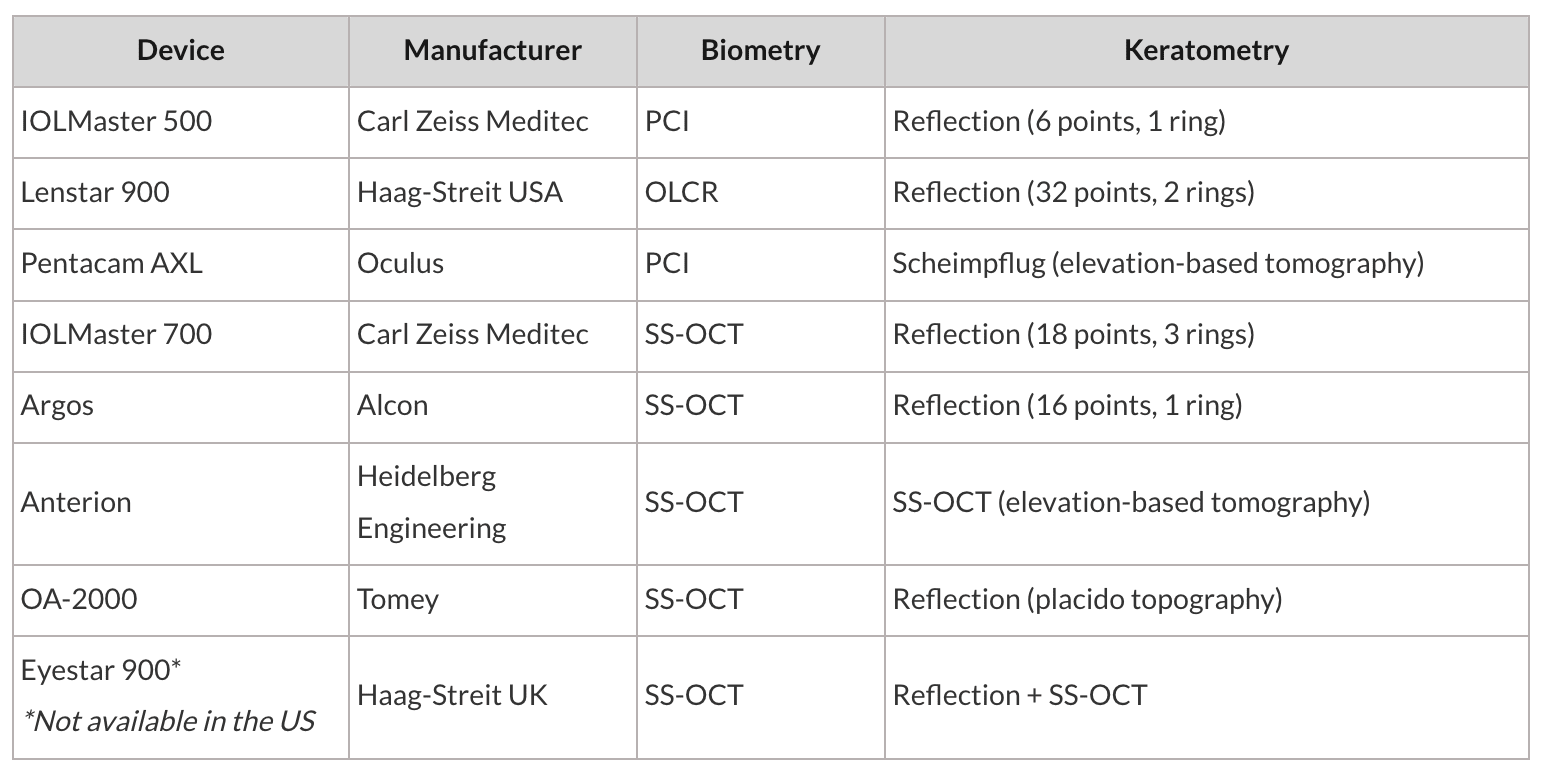
Since the introduction of the IOLMaster 500 (Carl Zeiss Meditec) in the year 2000, optical biometry has largely replaced U/S biometry as the preferred method for ocular measurement.6 Optical biometry relies on the principle of interferometry to measure the different segments of the eye, producing much higher resolutions of ±0.02 mm.7 In addition to its high resolution, optical biometry provides patients with a fixation target to ensure measurements correspond with their fovea. These optical measurements of the eye must be converted to physical distances.8 In the original device, this was accomplished through regression fit to immersion U/S data.2
As with any technology, optical biometry has seen substantial evolution over the last 25 years, transitioning from partial coherence interferometry (PCI) through optical low-coherence reflectometry (OCLR) to swept-source optical coherence tomography (SS-OCT). This evolution has resulted in improved tissue penetration, faster acquisition rates, higher resolution, and enhanced visualization. Interestingly, despite these noted improvements, measurements produced by all subsequent generations have been regression fit to prior generations, tracing all the way back to immersion U/S.
Historically, patients with mature cataracts often required U/S biometry because optical methods could not penetrate dense lens tissue.9 Advances in optical biometry, specifically SS-OCT, have substantially reduced this risk, with current optical biometers approaching 98% success.10 Despite this high success rate, surgeons will still encounter lenses that can only be measured with U/S, so U/S biometry remains an integral component of our diagnostic suite. If optical methods do fail, pharmacologic pupil dilation may facilitate AL acquisition in some patients with dense cataracts.11
Keratometry can be performed directly with optical biometers. These “auto-Ks” are generally reflection-based with resolutions comparable to manual keratometry and are measured in a smaller central zone of approximately 2.3 mm.12 Advancements in anterior segment imaging have led to the adoption of Scheimpflug and OCT-based imaging in newer biometers. These techniques permit measurement of the posterior corneal curvature, which may provide added benefit in eyes following corneal refractive surgery or when toric IOLs are planned.
Pearls for Accurate Measurement and Interpretation
As we alluded to previously, meticulous attention to measurement data is key to maximizing the refractive outcomes of cataract surgery. Our recommendations for biometry and keratometry include the following:
-
Verify the biometric data is of acceptable quality. Each device will provide quality metrics, eg, standard deviations, and a data summary for each patient. A quick inspection of the data will tell you how much trust you can place in your measurements. Recommended standard deviations for AL, keratometry magnitude, and steep meridian location are ≤0.02 mm, 0.3 D, and 3.5°, respectively.
-
Investigate AL asymmetries between fellow eyes >0.2 mm. While up to 24% of patients will have AL asymmetries >0.3 mm, you should still look for an explanation, eg, a scleral buckle or more myopic prescription in the longer eye. If no explanation is found, consider repeating the measurements using the same or a different device.
-
Obtain corneal topographies or tomographies on every cataract patient. Assess overall corneal regularity and look for evidence of ocular surface disease, epithelial basement membrane dystrophy, nodular degeneration, ectasia, or prior undisclosed refractive surgery. Also, investigate asymmetry in both keratometry magnitudes and meridians, as most patients’ corneas will have astigmatism readings that are mirror images of each other.
-
Obtain biometry and keratometry prior to instilling any drops in the eye. If the patient has significant dryness, closing the eye for 5 minutes has been shown to increase the likelihood of acquiring acceptable corneal measurements and avoids the potential refractive power shifts seen with artificial tears.
-
Pay attention to the selected measurement mode, ie, phakic, pseudophakic, aphakic, silicone oil, etc., and determine if the ACD and LT are within the expected ranges of 2.5 to 4.5 mm and 4 to 5.5 mm, respectively. Take note, optical biometers may run into trouble identifying the posterior lens surface, which can affect modern IOL formulas that use LT in their calculations, such as the Barrett Universal II.
Other Considerations
All biometers will offer a selection of IOL calculation formulas for direct use within the device. In general, these will include legacy formulas, such as the Holladay 1, SRK/T, and Haigis, but may also include more modern formulas such as the Barrett Suite, Hill-RBF, or Kane. Built-in formulas avoid the risk of transcription errors, but are limited to those with whom manufacturers hold licensing agreements. Surgeons can always explore other web-based IOL calculation formulas when the clinical scenario calls for it.
Depending on the device manufacturer, biometers may offer additional levels of integration with other equipment involved in the cataract workflow. They may communicate directly with femtosecond lasers, phaco machines, operating microscopes, and/or surgical planning software. This automated transfer of data can provide several advantages for the cataract surgeon, such as intraoperative heads-up data display, toric IOL alignment, and outcomes analysis.
Conclusion
Patients and surgeons have certainly benefitted from the continual evolution of ocular biometry and keratometry, as well as the industry’s continued interest in furthering development in this field. Beyond improving accuracy, modern optical biometers can also enhance clinical efficiency and practice economics. Optical biometry requires less operator training and enables rapid acquisition of multiple measurements in a single examination, streamlining workflow and reducing repeat testing. Integration with various elements of the cataract workflow can minimize transcription errors, reduce administrative burden, and support IOL selection and postoperative outcomes tracking. Although advanced biometers require a substantial upfront investment, their high acquisition rates and reduced reliance on ultrasound can improve overall practice efficiency. When selecting a biometer, clinicians should consider not only its overall performance but also its individual features, cost, and integration into their specific practice workflow. OM
References
- Sahin A, Hamrah P. Clinically relevant biometry. Curr Opin Ophthalmol. 2012;23(1):47-53. doi:10.1097/ICU.0b013e32834cd63e
- Findl O, Drexler W, Menapace R, Heinzl H, Hitzenberger CK, Fercher AF. Improved prediction of intraocular lens power using partial coherence interferometry. J Cataract Refract Surg. 2001;27(6):861-867. doi:10.1016/s0886-3350(00)00699-4
- Shammas MC, Shammas HJ. Ultrasound biometry. In: Aramberri J, Hoffer KJ, Olsen T, Savini G, Shammas HJ, eds. Intraocular Lens Calculations. Essentials in Ophthalmology. Springer; 2024:163-175. doi:10.1007/978-3-031-50666-6_9
- Gurnani B, Kaur K. Keratometer. In: Khandpur RS, ed. Compendium of Biomedical Instrumentation. John Wiley & Sons; 2023:1109-1112. doi:10.1002/9781119288190.ch206
- Friedman NJ, Kaiser PK. Optics/refraction. In: Case Reviews in Ophthalmology. 2nd ed. Elsevier; 2018:1-46. doi:10.1016/B978-0-323-39059-0.00001-0
- Miller KM, Oetting TA, Tweeten JP, et al. Cataract in the adult eye preferred practice pattern. Ophthalmology. 2022;129(1):P1-P126. doi:10.1016/j.ophtha.2021.10.006
- Khorrami-Nejad M, Khodair AM, Khodaparast M, Babapour Mofrad F, Dehghanian Nasrabadi F. Comparison of the ocular ultrasonic and optical biometry devices in the different quality measurements. J Optom. 2023;16(4):284-295. doi:10.1016/j.optom.2023.05.001
- Sen S, Tripathy K. Ultrasound Biometry. StatPearls. January 11, 2024. Accessed July 8, 2026. https://www.ncbi.nlm.nih.gov/sites/books/NBK599551/
- Mylonas G, Sacu S, Buehl W, Ritter M, Georgopoulos M, Schmidt-Erfurth U. Performance of three biometry devices in patients with different grades of age-related cataract. Acta Ophthalmol. 2011;89(3):e237-e241. doi:10.1111/j.1755-3768.2010.02042.x
- Akman A, Asena L, Güngör SG. Evaluation and comparison of the new swept source OCT-based IOLMaster 700 with the IOLMaster 500. Br J Ophthalmol. 2016;100(9):1201-1205. doi:10.1136/bjophthalmol-2015-307779
- Bettach E, Totah H, Weill Y, Zadok D, Abulafia A. Optimizing axial length measurement success in advanced cataract patients through pupil dilation. Indian J Ophthalmol. 2024;72(Suppl 5):S870-S874. doi:10.4103/IJO.IJO_3316_23
- Lin HY, Chen HY, Fam HB, Chuang YJ, Yeoh R, Lin PJ. Comparison of corneal power obtained from VERION image-guided surgery system and four other devices. Clin Ophthalmol. 2017;11:1291-1299. Published 2017 Jul 12. doi:10.2147/OPTH.S137878