

At the American Academy of Ophthalmology 2026 meeting in New Orleans, Neal H. Shorstein, MD, Naveen S. Chandra, MD, Gladys Lucia Silva Linares, MD, and Alastair James Stuart, BMBS, BMED, FRCOphth, discussed how a pair of surgeon-placed injections of intracameral moxifloxacin and subconjunctival triamcinolone acetonide provides a safe and effective alternative to a perioperative course of patient-instilled topical antibiotic, steroid, and NSAID drops.
“Patients have difficulty administering their own eye drops, especially the elderly, and more so in the postoperative period,” explained Dr. Chandra. “There's a risk of injury and contamination of the surgical wound when self-administering medications.So, the challenge we're trying to address is: how can we ensure good infection prophylaxis and inflammation control in cataract surgery?”
In the course, "Dropless Cataract Surgery: How to Get It Going in Your Practice,"
Drs. Shorstein and Chandra highlighted results from a study they conducted with Shannon E. McCabe, MD, Mubarika Alavi, MS, and Marilyn L. Kwan, PhD, titled “Triamcinolone Acetonide Subconjunctival Injection as Stand-alone Inflammation Prophylaxis after Phacoemulsification Cataract Surgery.” The retrospective, comparative effectiveness cohort study examined the safety and effectiveness of triamcinolone subconjunctival injection as prophylaxis during cataract surgery in patients at Kaiser Permanente Northern California from 2018 through 2021.
“We looked at our cataract surgery data to determine if there is an ideal dose and concentration of triamcinolone,” said Dr. Chandra. “After reviewing these cases for the inclusion criteria—not having other prior surgeries, not having a chronic condition like uveitis—we ended up with a population of about 69,800 eyes in our data set."
Exposure groups included topical prednisolone acetate (PA) with or without NSAID as well as subconjunctival injection of triamcinolone acetonide (Kenalog; Bristol-Myers-Squibb) 10 mg/mL or 40 mg/mL in a low dose (1.0-3.0 mg) or high dose (3.1-5.0 mg) (Figure 1).
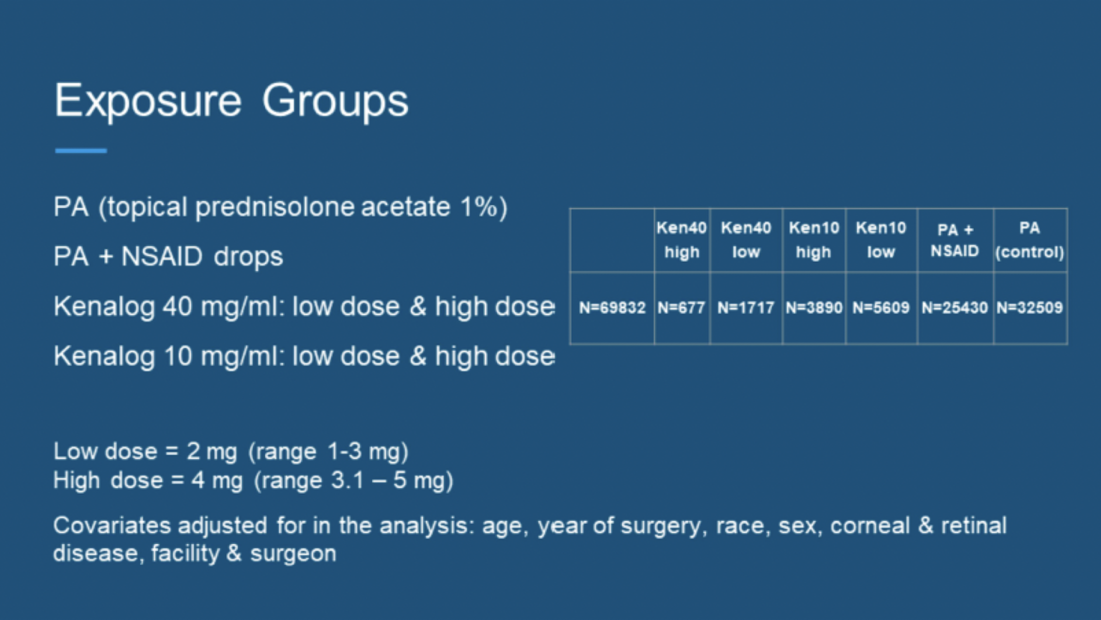
"When comparing these groups, we measured effectiveness by determining the rates of postoperative macular edema and iritis, while we used the rate of glaucoma events to evaluate the safety of the steroid injection," noted Dr. Chandra.
The study concluded that the Kenalog 10 mg/mL high-dose injection group (~4 mg) had 36% lower odds of postoperative macular edema, similar odds of a glaucoma event, and similar odds of rebound iritis when compared with topical PA. Prednisolone and NSAID were comparable to prednisolone alone—thus Kenalog 10 mg/mL injection alone is sufficient in controlling inflammation. In Kenalog 40 mg/mL groups, there were statistically significant higher odds of a glaucoma event.
With these results, Dr. Shorstein and Dr. Chandra recommended the following practice pattern to their peers within Kaiser Permanente (Figure 2).
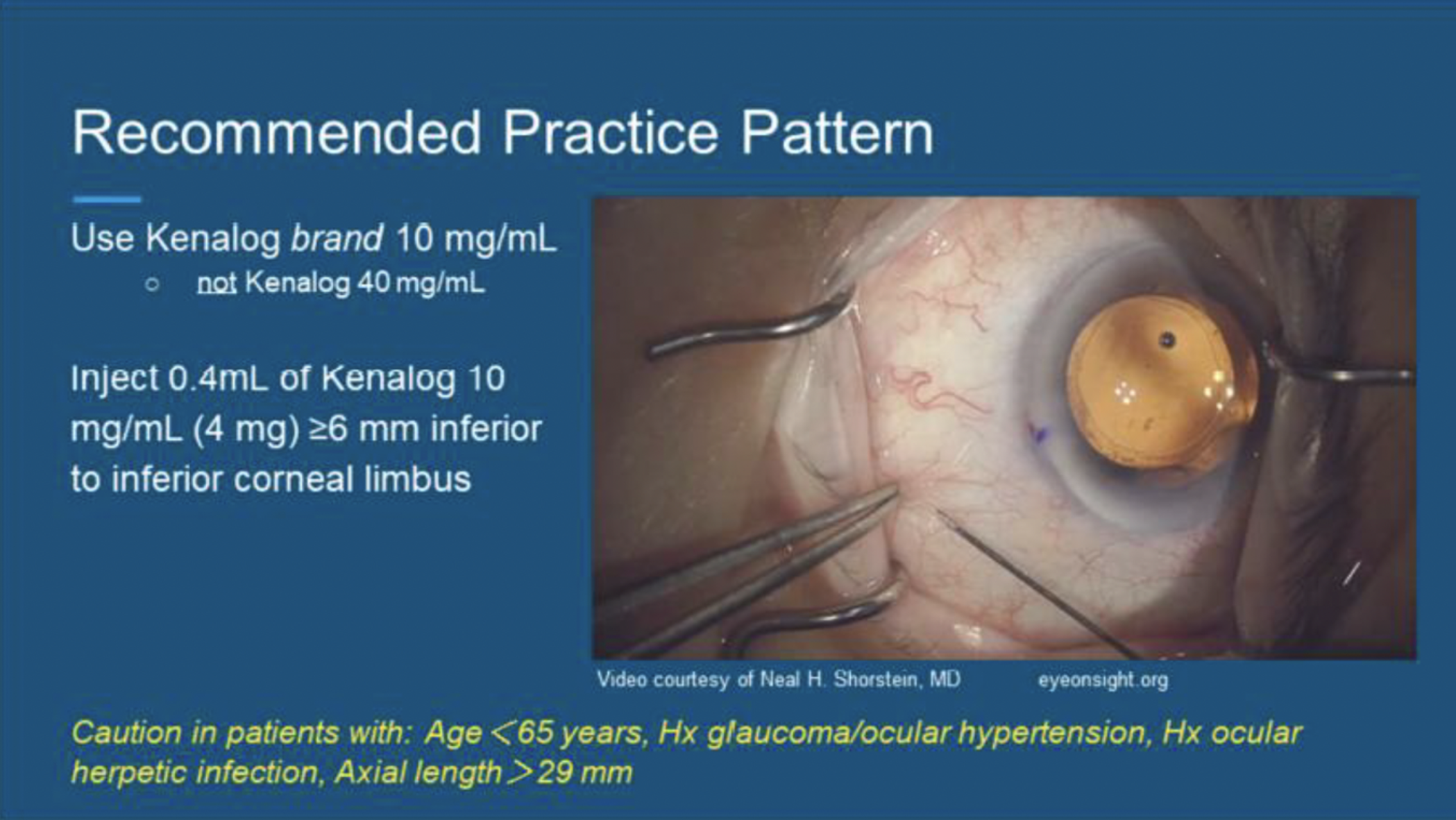
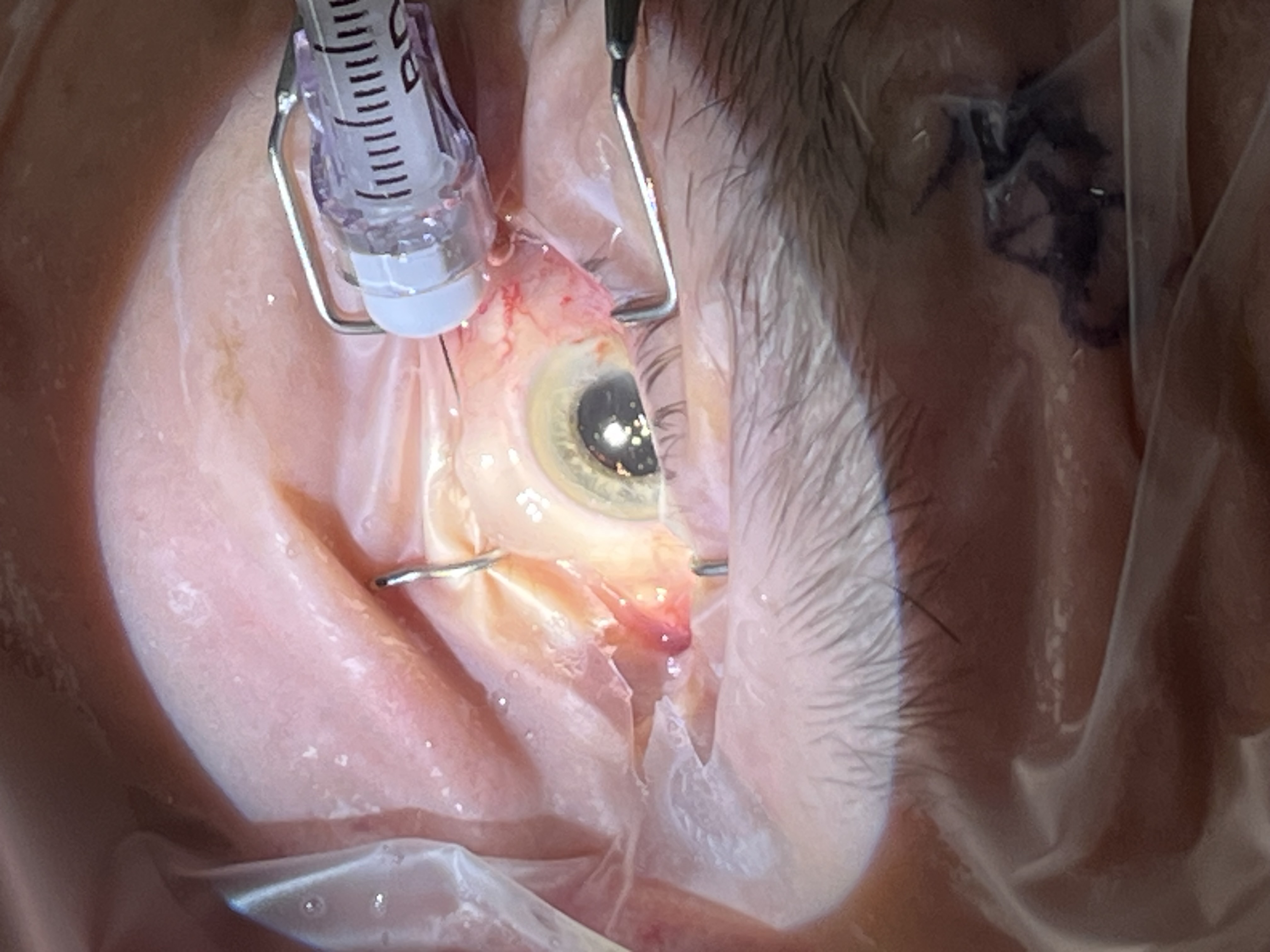
“We recommended using Kenalog brand 10 mg/mL—not Kenalog 40 mg/mL—and to inject 0.4 mL of Kenalog 10 mg/mL (4 mg) 6-8 mm inferior to the inferior corneal limbus,” said Dr. Chandra. “Our data are specific to brand-name Kenalog, not as a brand endorsement, but because each brand has different particle shape and size, so each brand exhibits different pharmacodynamics" (Figure 3).
However, he stressed caution in patients who are <65 years old, have a history of glaucoma/ocular hypertension or ocular herpetic infection, and those with an axial length of >29 mm.
“Patients with these characteristics have been shown to be at increased risk of intraocular pressure spikes with steroid treatment," he explained.
Dr. Chandra concluded, "I think the take-home message from our experience is that subconjunctival steroid injection at the time of cataract surgery is a safe and effective way to address postoperative inflammation. This method has been extremely popular with patients given that it removes the burden of self-administering eye drops. The use of intracameral antibiotic as infection prophylaxis is standard-of-care, and I encourage surgeons to consider adopting this dropless method of inflammation prophylaxis to accompany it.”
He continued, “In addition, by administering necessary postoperative medications intraoperatively, we can reduce the hassle as well as the carbon footprint of patients and family members having to collect medications from pharmacies.”
Dropless Is Sustainable
Dr. Stuart added that dropless surgery also offers sustainable advantages. For example, he noted, one intraoperative depot can replace a multiweek course of postoperative steroid drops. Dropless surgery uses less material—with fewer eye-drop bottles, cartons, leaflets, and single-use packaging. Dropless surgery also creates a leaner supply chain with less prescribing, dispensing, storage, and movement of medicines. Less waste is another advantage, with fewer partly used or unused bottles entering clinical or household waste, he said.
“Dropless is primarily a patient-care innovation, but reducing the postoperative medication pathway also removes avoidable resource use,” he explained (Figure 4).
Dr. Stuart also presented findings from a study on dropless cataract surgery that was conducted at Optegra Eye Health Care, which included more than 15,000 patients. “As the idea of dropless cataract surgery is increasing in popularity, it is important for clinicians to follow published evidence,” he emphasized.
Dr. Stuart explained that in the head-to-head dropless vs standard postoperative regimens, the standard protocol included a local anesthetic; standard phacoemulsification and lens implantation; and dexamethasone ophthalmic suspension 0.1% (Maxidex; Harrow) plus intracameral cefuroxime. The dropless protocol included local anesthetic; standard phacoemulsification and lens implantation; and intraoperative subconjunctival triamcinolone 0.4 mL of 10 mg/mL plus intracameral cefuroxime. The investigators compared 30,000 dropless procedures with the original drop regimen.
Regarding cystoid macular edema, there was no statistically significant difference between pathways (all P values >.05), with 3.1% (dropless pathway) vs 3.3% (drop pathway). Regarding intraocular pressure spikes, Dr. Stuart noted equivocal results between pathways, with no additional pressure risk from the dropless regimen: 0.60% (dropless pathway) vs 0.68% (drop pathway).
“We have shown the results in terms of visual outcomes and complications to be equivocal to surgery with traditional drops,” he said. “The benefits are increased convenience and experience for the patient, and due to reduced packaging, there is a significant sustainability improvement.”